


Third Occipital Nerve Block and Radiofrequency Neurotomy
What is third occipital nerve block? Why is it done?
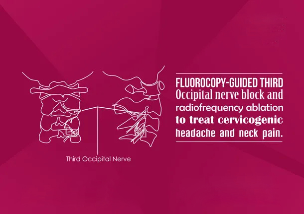
Third occipital nerve supplies the pair of tiny joints present higher up in the neck, closer to the skull called the C2/3 facet joint, and a small part of skin below the occiput (a raised prominence in the back of the skull). The third occipital nerve block is performed to relieve headache caused by the degeneration of the facet joint known as cervicogenic headaches. This headache typically originates at the back of the head and extends over the top of the head on one side. It is usually associated with arthritic inflammation of the C2/C3 joint or inflammation of the third occipital nerve itself.
How is it done?
At Atlas Pain Care, Coimbatore, you will be asked to change into a hospital gown and taken to our procedure room. In the procedure room you will be asked to lie face down on a cushioned X-ray table. The back of your neck will be cleaned with antiseptic solution and draped. The injection site is then identified using X-rays and a small amount of local anaesthetic is injected into the skin overlying the injection site to numb the skin. The medications are then given through a longer needle that has been positioned in the exact spot using ultrasound guidance. You may feel some discomfort during the injection, but this normally settles quickly. The whole procedure will take around about 15 minutes, and post procedure you will be observed in recovery room for 30 minutes and then discharged home. You may need to rest at home for the remainder of the day. But you should be able to resume your normal activities the next day.
What is radiofrequency ablation of third occipital nerve?
If the patient gets good pain relief for few days with third occipital nerve block then a radiofrequency lesion is proposed. In this, along with the X-ray, an electric needle is used to identify the nerves. The nerves are then stunned with radiofrequency current to provide long-term benefit.
What are the possible risks?
Complications are minimal as these injections are done under image guidance. Pain in the area where the needle was inserted can last for two to three days. Bruising at the site of infection can occur. Other complications like infection, bleeding and nerve injury are very rare. Please read our FAQ section to know more about the procedure.